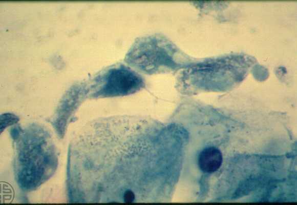
Trichomonas vaginalis
Trichomonas vaginalis. Trophozoites
in vaginal smear (Papanicolaou stain, oil immersion). Several trophozoites are
seen to be linearly arranged in this field. The middle organism that stains
more darkly demonstrates a fine flagellum extending from it. Two of the trophozoites
to the right of this flagellated trophozoite demonstrate characteristic red
granules (hydrogenosomes), whereas they are less clear in the other organisms.
The variable morphology and size of these trophozoites illustrate the difficulty
in recognizing them in Papanicolaou stained smears.