Microsporidians
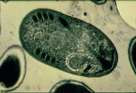
Encephalitozoon hellem. Spore
in tissue section (electron microscopy). A mature spore of E. hellem
demonstrates the typical morphology of a microsporidian spore when viewed with
transmission electron microscopy. The sections of the coiled polar tubule seen
as peripheral black structures on both sides at the periphery of the spore wall
rapidly identify the spore as that of a microsporidian.